Upstate introduces new surgical navigation system for complex endovascular cases
Upstate Medical University’s Heart and Vascular Center recently implemented a cutting-edge GPS-like surgical navigation device for use in complicated cases.
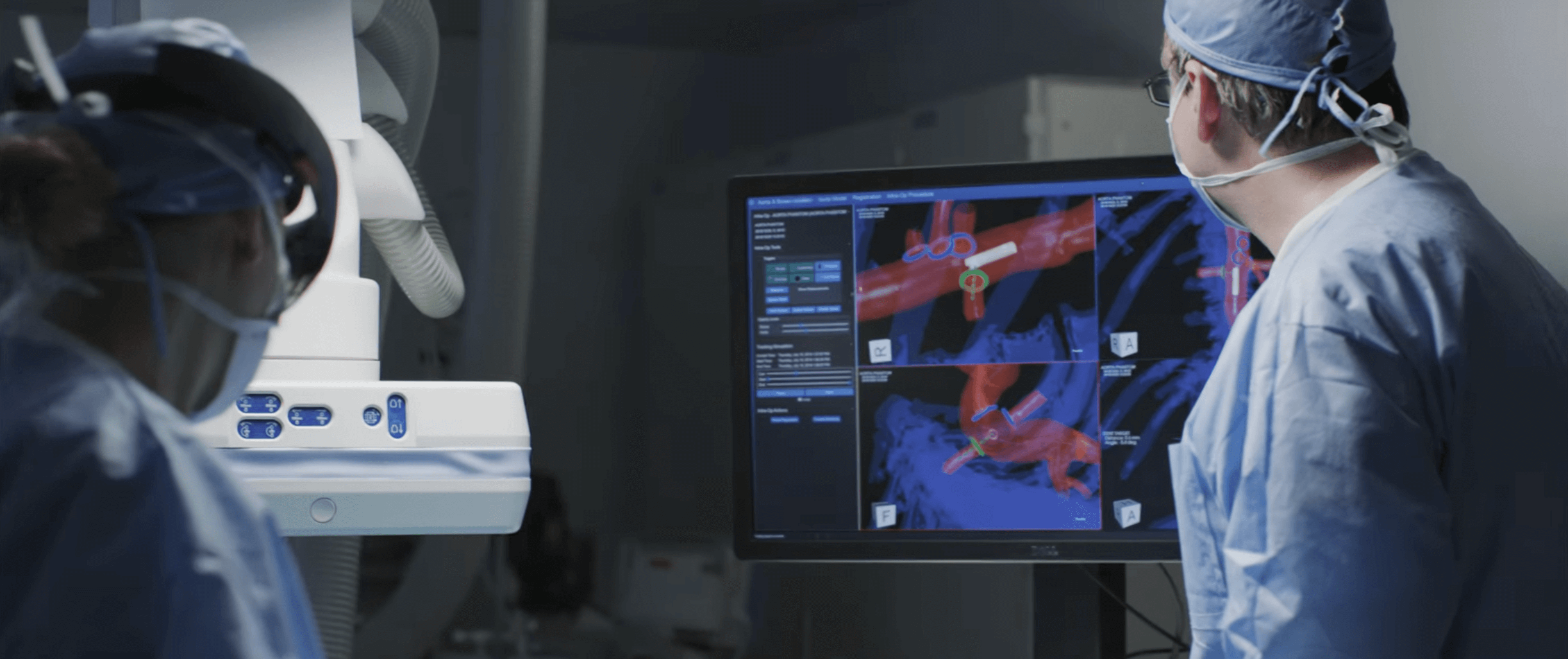
The Inter-Operative Position System (IOPS) offers doctors better views and greater control during complex endovascular procedures while reducing the need for X-rays. It was used for the first time last month to easily insert a stent into blood vessel that’s normally hard to see and access.
The surgery was performed by Michael Costanza, MD, who said the surgery was needed to increase blood flow to the patient’s intestines after he suffered an aortic dissection. The blood vessel in question is at a difficult angle to access and it overlaps another vessel so it’s hard to see on X-ray, which is what is normally used to guide these types of procedures.
With the IOPS, Costanza was able to place the catheter in less than a minute. Using X-ray, that could have taken up to 30 minutes.
“The new system makes it so straightforward,” said Costanza, division chief of Vascular Surgery and Endovascular Services. “You’re getting to where you need to go accurately; it’s very reassuring.”
Senior radiology technician James Glowacki said the IOPS is a mapping system that uses magnetic fields. Once calibrated to the patient on the table, doctors can view the inside of the patient’s body in 3D.
Patients first get a CT scan, and that image is combined with the IOPS system for a full view of their body that can be manipulated to see any angle necessary. It will be used for complicated procedures such as repairing abdominal aortic aneurysms or thoracic aortic aneurysms.
“This technology improves patient outcomes, decreases radiation exposure, and shortens the duration of the procedure,” Costanza said.
Upstate is just the 10th hospital in the country to use the navigation system and the first in the Northeast.
Without this system, X-rays are used during the procedure to help guide the doctor, but those provide only two-dimensional images. Typically, X-rays are used for much of the duration of these surgeries.
With the IOPS, doctors can see the catheter and wire travel through the blood vessel in real time 3D. Doctors see everything on a big monitor.
“Not only do you see that patient’s anatomy, but you also see your tools,” Costanza said. “You see your catheter and your wire in there. It’s like a video game. It’s amazing technology.”
According to Centerline Biomedical, which makes the IOPS, complicated endovascular cases have a 15 times higher dose of radiation as compared to a standard one. The IOPS reduces time needed to cannulate a vessel by almost 50 percent and completely eliminates the need for X-rays.
Heart and Vascular Center Director Amy Tetrault said the system can lead to improved outcomes for patients because doctors can complete the surgery in less time. She said a three- or four-hour surgery could be shortened an hour using the IOPS.
“It will be great for our patients,” Tetrault said. “It shows the dedication of our physicians to want to bring this technology here to Upstate and to our patients. We are excited to see its benefits.”
Costanza said the technology is already evolving and he has seen prototypes of the next step forward.
“The next generation of this will be using VR glasses that allow you to just look at the patient,” he said. “Everything will be projected right on the patient. It’s like science fiction but that’s already being developed.”
The Heart and Vascular Center used the IOPS twice more in February for a splenic artery aneurysm and for a celiac artery occlusion.

