IOPS® Software
IOPS Software Key Features
IOPS (Intra-Operative Positioning System) is a 3D endovascular visualization and navigation tool designed to minimize radiation exposure and promote successful interventions
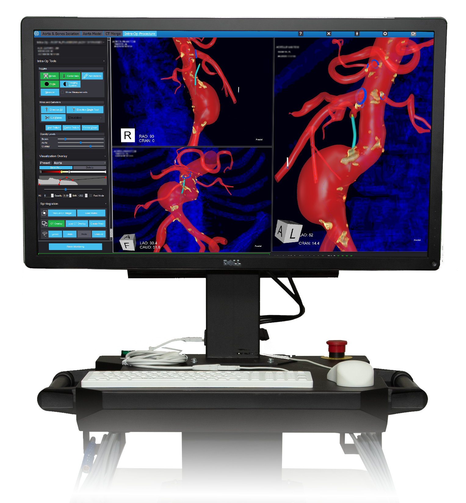
Build 3D, multi-color models of unique patient anatomy from existing CT scans

Show up to four views of the
patient anatomy concurrently

Capture 3D intraoperative image
updates with Spintegration™

Displays position and orientation
of IOPS Catheters & Guidewires,
and implanted devices

Works with all
CBCT imaging systems

Rotate, magnify, and annotate
procedural targets within the
3D model in real time

Build 3D, multi-color models of unique patient anatomy from existing CT scans

Show up to four views of the patient anatomy concurrently

Displays position and orientation of IOPS Catheters & Guidewires, and implanted devices


New to IOPS Software
Version 1.5 empowers clinicians with more ways to reenvision endovascular navigation
Enables the Use of 6Fr Viewpoint™ Catheters & New Accessories
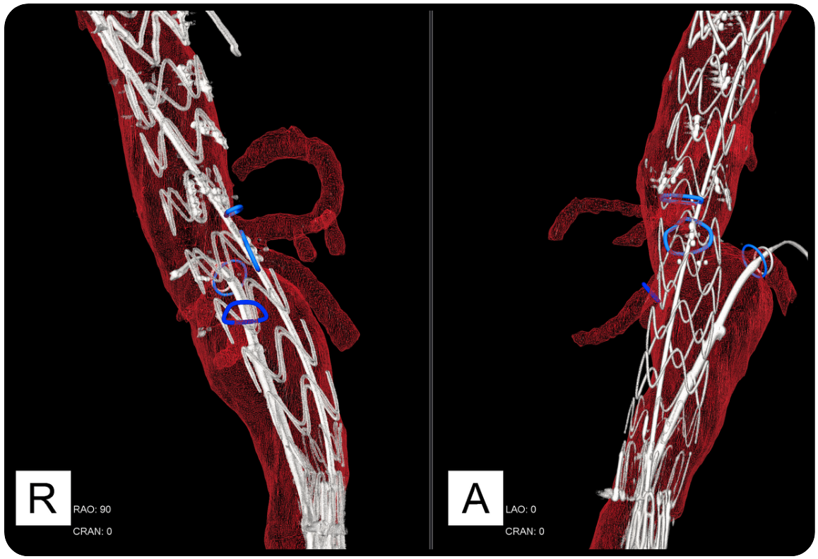
3D Mesh Wireframe
Vessel Display
Improves Model Development, Vessel Isolation, and Annotations
Biplane Lock and Enhanced Encrypted DICOM Data Transfer
Updates 3D Modeling
Capabilities for CT Snapshots
Additional User Interface Enhancements
Enhanced visualization displays fine anatomical features
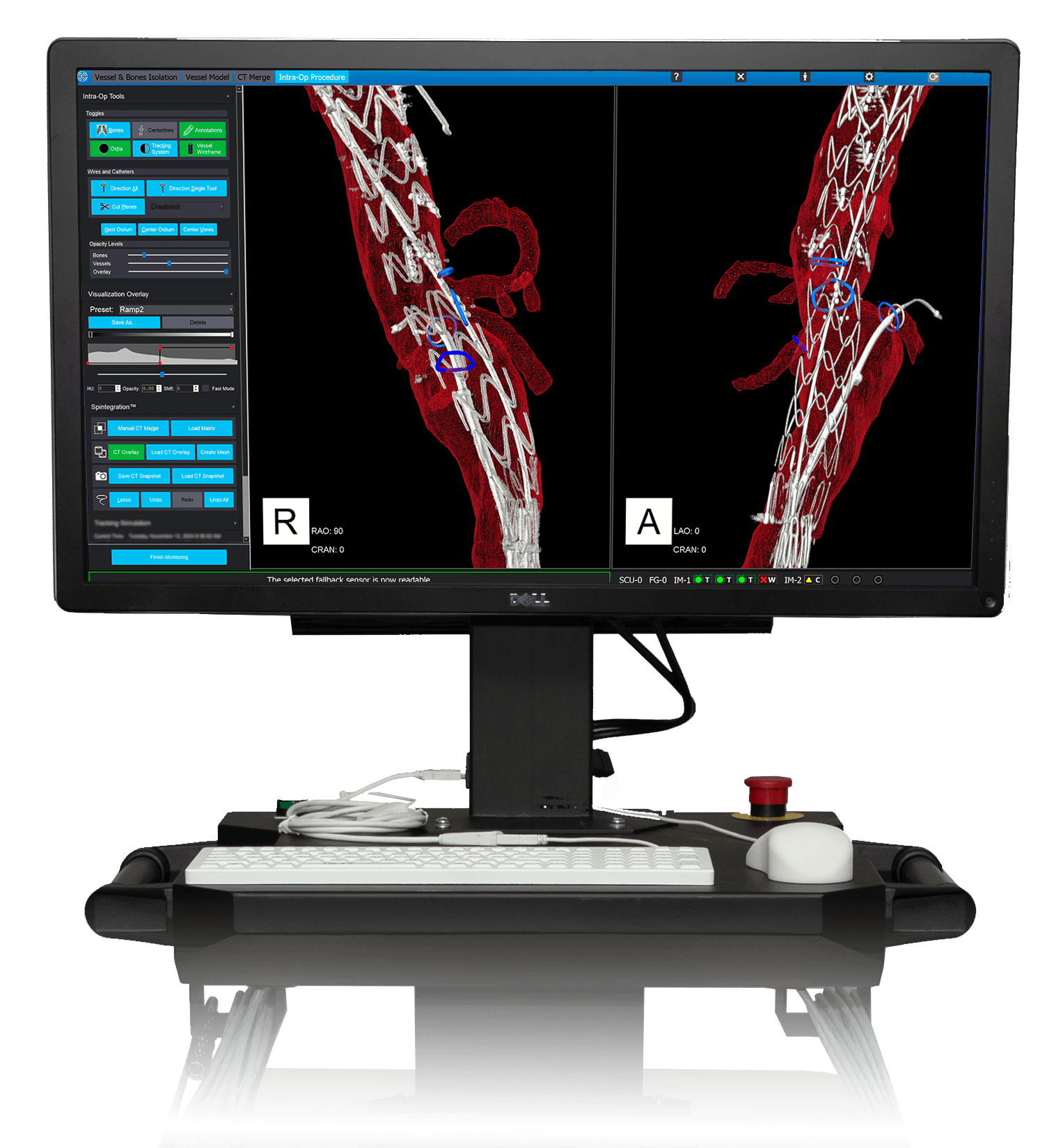
3D mesh wireframe vessel display
IOPS Software Version History
- Enables the use of 6Fr Viewpoint™ Catheters and new accessories
- Biplane Lock feature automatically syncs multiple projections
- Adds support for encrypted DICOM data transfers
- CT Snapshot enhances case planning, mapping, and visualization of calcifications, endografts, and endoleaks
- Adds 3D mesh wireframe vessel display
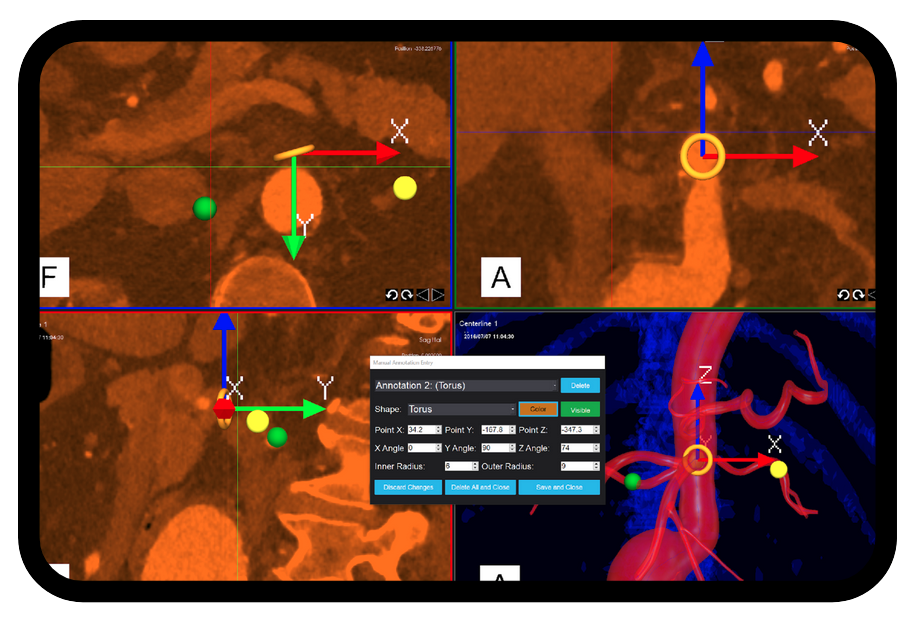
- Adds ability to place annotations in any workflow
- Spintegration enhancements: Improves ability to focus on navigation targets. Improves ability to visualize unique patient anatomy.
- Customizes color selection based upon clincian preference
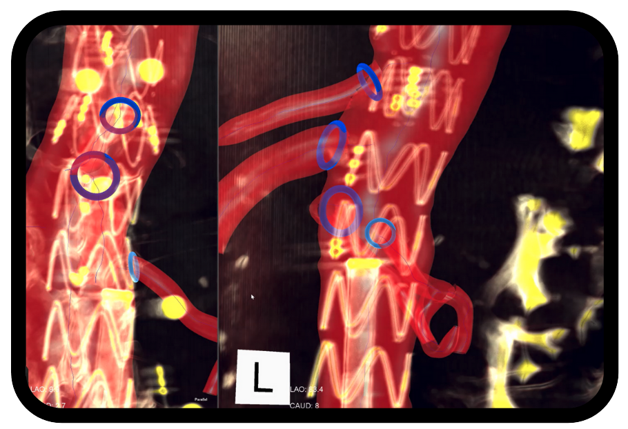
- Introduction of Spintegration to enable display of CBCT scan overlay in Intra-Op workflow
- Adds ability to show deployed stents and other implantable devices
- Automates and simplifies model building from vessel isolation
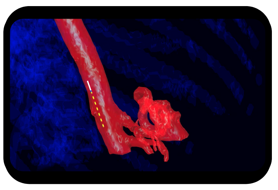
- Expands directional indicator feature, available on IOPS Catheters
- Supports extending visual output of up to four views on external OR monitors
- Performance improvements on model building and vessel isolation
- Aligns IOPS view with fluoro views
- Improves visual confirmation of tool connectivity
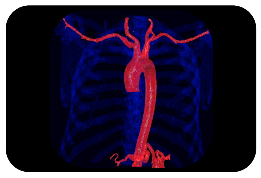
- Improves model building for smaller vessels and the branching structure
- Adds performance support for multiple cathethers in complex views
- Adds en face and on edge views of ostium
- Adds screen capture and recording support
- Compatibility with more accurate, next generation field generator and sensor control unit
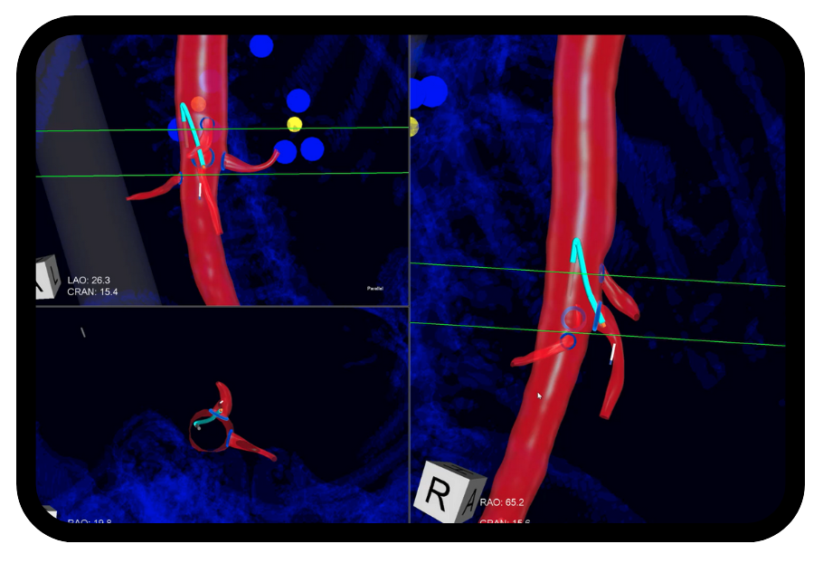
- Build 3D, multi-color models of unique patient anatomy from existing scans
- Provides real-time tip positioning and navigation using sensor equipped compatible catheters and guidewires
- Rotates 3D models of the patients anatomy in real time
- Shows up to four 3D views of the patient anatomy concurrently
- Compatible with all CBCT imaging systems; including GE, Siemens, and Philips*
*All trademarks are the property of their respective owners
Learn how IOPS can positively
impact your workflow:
